
Ocular Dermoid Cyst in a neonate Calf
A historical entry from the OASIS animal picture blog. Original observations and discussion are preserved; this entry has not received a new veterinary review. Some photographs show clinical procedures.
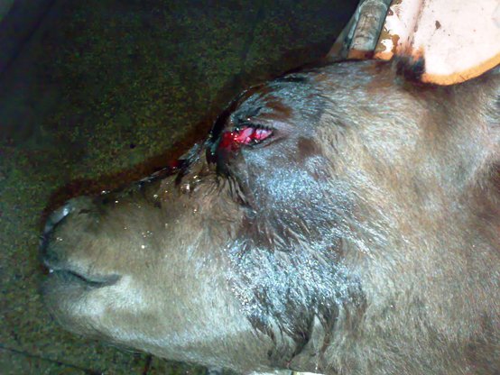
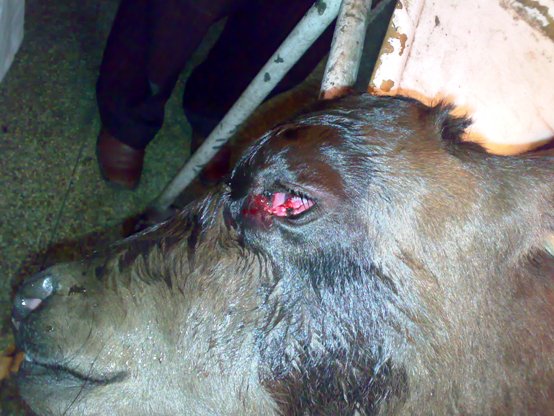
A day-old calf was presented at the veterinary clinic with bilateral corneo-scleral dermoid cysts which were completely occluding the lid opening and rendering the calf blind. The cysts extended from the cornea , partially covering it; and travelling medially fusing with the third eye lid.
A surgical procedure was attempted on the calf with the aim to one, remove as much of the mass of the cyst as possible so as to remove the obstruction to light and second. to peel off the dermal layers containing the follicles as close as possible to the cornea/sclera so as to prevent regrowth of hair follicles. Sutures were placed to promote fusing of the raw tissues with the lower eyelid.
The first four pictures show the extent of the cyst and occlusion due to it
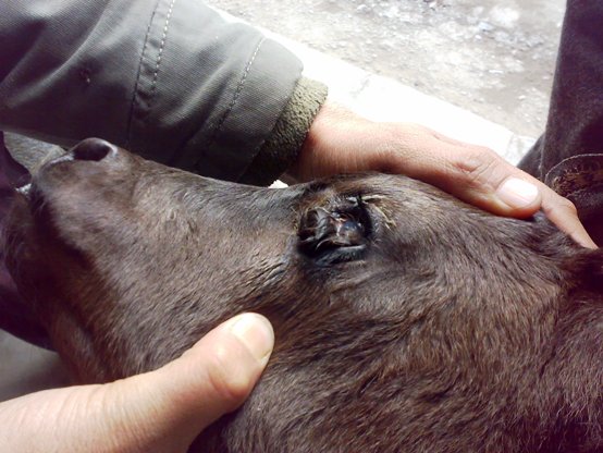
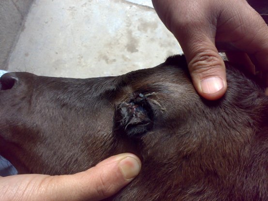
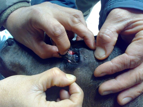
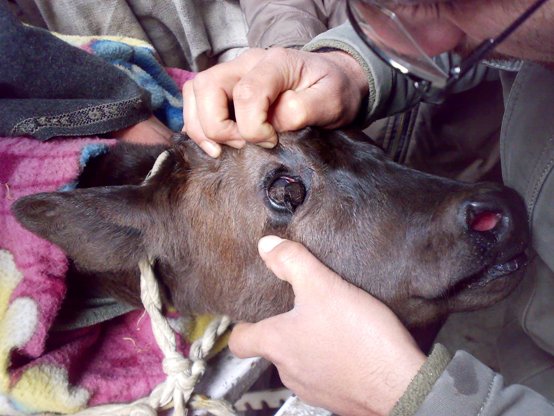
Sutures were placed through the tissue buldge and later the tissue excised.
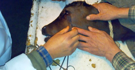
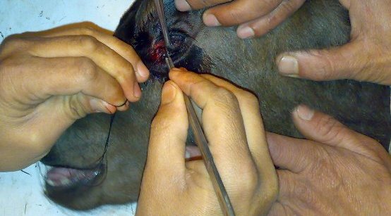
The upper layers of the skin are sliced through to promote fibrosis and prevent hair growth.
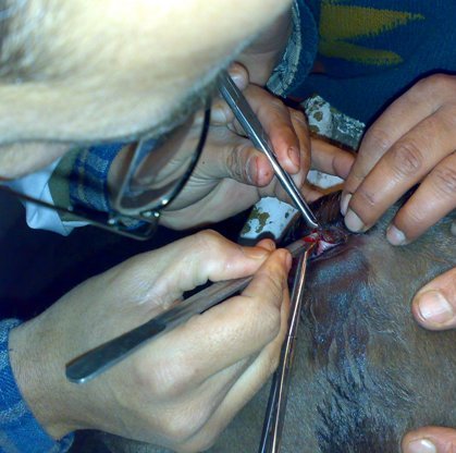
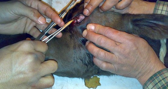
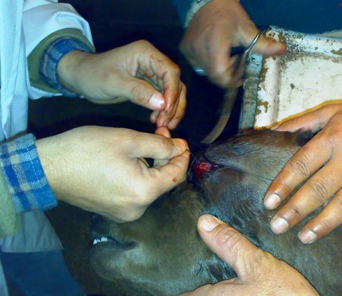
Finally an almost proper opening between eyelids
From the original discussion
Archived comments are preserved in their original context. New comments are closed.
Dear dr. anjum
As always I appreciate your concern for the veterinary profession.The dermoid pictures are of good quality however the basic tenets of Halstead taught to every graduating veterinarian in first few classes of surgery have to be followed at all costs.Most of them are practicable even field.Your pictures are being viewed throughout world.
I assure you that your advice has been taken and will be followed strictly..