By Anjum Andrabi · Originally published September 3, 2008
A historical entry from the OASIS animal picture blog. Original observations and discussion are preserved; this entry has not received a new veterinary review. Some photographs show clinical procedures.
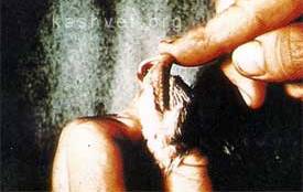
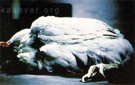
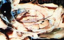
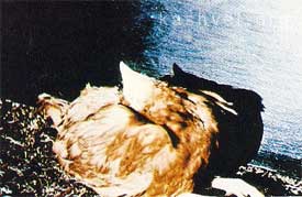
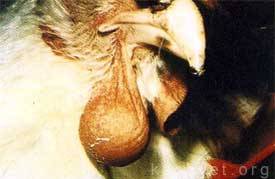
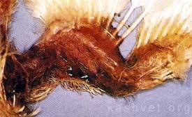
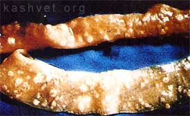
Decreased mineralization of the mandible and maxilla allows extensive lateral movement of the beakHen showing sternal recumbancy due to osteomalaciaSevere nephritis characterised by pallor and urate deposition may be associated with nephrogenic strains of infectious bronchitis, infectious bursal disease or water deprivationTorticollis may be due to infection of the inner ear by E. coli or Pasteurella multocida. It has also been observed in flocks suffering from velogenic Newcastle diseaseEnlargement of wattle due to Pasteurella multocida infectionWing of 14-day old broiler showing edema, subcutaneous hemorrhage and transudate due to chick anemia virusMucosa of intestine showing lesions characteristic of necrotic enteritis
From the original discussion
Archived comments are preserved in their original context. New comments are closed.

From the original discussion
Archived comments are preserved in their original context. New comments are closed.
hello
respectfully i need a collection of gross lessines in all of poultry diseases thanks