By Anjum Andrabi · Originally published September 2, 2008
A historical entry from the OASIS animal picture blog. Original observations and discussion are preserved; this entry has not received a new veterinary review. Some photographs show clinical procedures.
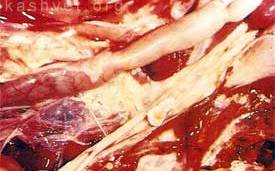
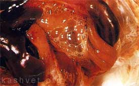
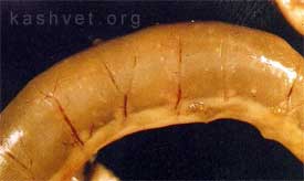
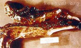
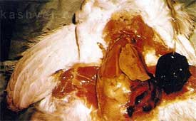
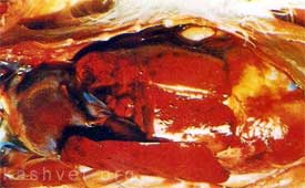
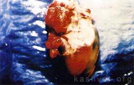
Caseous airsacculitis associated with Mycoplasma gallisepticum infectionAcute foamy airsacculitis due to Mycoplasma gallisepticum infectionEimeria acervulina/mivati infection showing white foci visible through the serosa of the duodenumSevere Eimeria tenella infection showing hemorrhagic cecaFatty liver syndrome in mature hens may result in rupture and hemorrhage into the body cavityCharacteristic mottled appearance of liver (subcapsular hemorrhage) associated with Adenoviral hepatitis infection and specifically HHS (Leechi disease)Characteristic hydropericardium associated with Angara disease (Leechi disease)