By Anjum Andrabi · Originally published September 2, 2008
A historical entry from the OASIS animal picture blog. Original observations and discussion are preserved; this entry has not received a new veterinary review. Some photographs show clinical procedures.
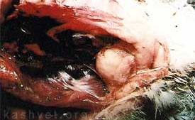
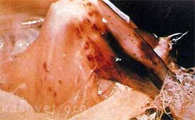
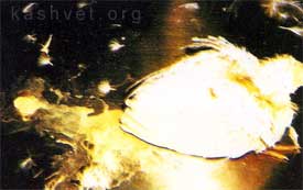
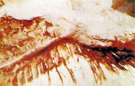
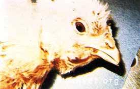
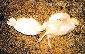
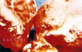
Enlarged Bursa of Fabricius typical of acute infectious bursal disease. Note urate accumulation on vent plumage and enlarged kidneysCharacteristic subcutaneous and intramuscular hemorrhages associated with infectious bursal diseaseAcute infectious bursal disease showing chicken with copious white diarrheaHaemorrhage and transudate beneath the wing associated with transduative diathesis due to destruction of Vitamin E following oxidative rancidityChicks showing edema of intamandibular region due to transduative diathesisBroiler chicks approximately 14-days old showing lateral recumbency associated with nutritional encephalomalacia Hemorrhage within the cerebellum characteristic of nutritional enchepalomalacia due to Vitamin E deficiency